Photo courtesy of Artemii Savin
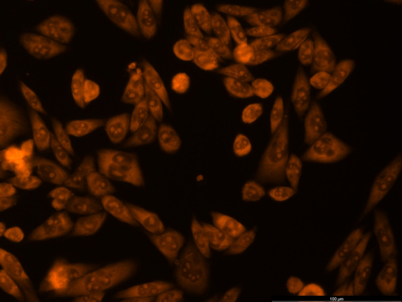
The orange objects in the picture are cells of human neuroblastoma, a malignant tumor that develops from immature nerve cells.
Neuroblastoma interests scientists not only because its in-depth study might help them fight this type of cancer. The location of human neurons – in the brain and spinal cord – makes them unavailable for direct scientific study. Therefore, neuroblastoma cells can serve as an experimental model of human neurons and help analyze drugs aimed at treating nervous diseases.
Before embarking on expensive human or animal trials, the efficacy and safety of any drug need to be assessed. As healthy brain cells are unsuitable for this research, tumor cells that have the same basic components as typical neurons can partially be used for this purpose.
In the picture, living cells are depicted in red (stained with acridine orange). The picture was taken with a Leica DMi8 fluorescence microscope in the rhodamine channel and allowed the scientists to assess the viability and state of cells. The experiment was carried out by Master’s students Artemii Savin and Anastasiya Kryuchkova as part of a joint project (Experimental Oncology and Immunology and Ceramic and Natural Nanomaterials groups).
(source https://news.itmo.ru/en/science/life_science/news/9937/)